SUSPECTED HYDRATED NUCLEUS PULPOSUS EXTRUSIONS ASSOCIATED WITH DORSAL LONGITUDINAL LIGAMENT IN DOGS
Background
Disc-associated intraspinal cysts and cervical hydrated nucleus pulposus extrusions are lesions of suspected disc origin recently described in veterinary literature.
Introduction/Purpose
To describe MRI, surgical and cytological findings in patients with acute hydrated cervical disc extrusions associated with the dorsal longitudinal ligament (DLL).
To describe MRI, surgical and cytological findings in patients with acute hydrated cervical disc extrusions associated with the dorsal longitudinal ligament (DLL).
Material and Methods
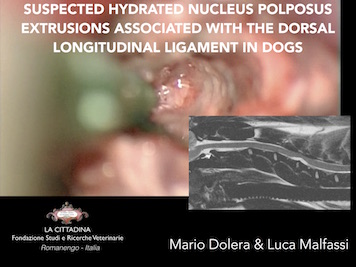
Prospective study of dogs with evidence of cervical spinal cord compression by material associated with the dorsal longitudinal ligament (DLL). Inclusion criteria were: availability of high-field MRI examination, microsurgical decompression and cytological examination of the removed material. The following MRI features were assessed: grading of spinal cord compression, intervertebral spaces involved, signal intensity and distribution of the material, T2-W spinal cord signal, thickness and signal intensity of the discs. Spinal cord compression was evaluated in cross-sectional T2-W images. Surgical findings considered were: macroscopic appearance of the material, its localization and relationship with the DLL.
Prospective study of dogs with evidence of cervical spinal cord compression by material associated with the dorsal longitudinal ligament (DLL). Inclusion criteria were: availability of high-field MRI examination, microsurgical decompression and cytological examination of the removed material. The following MRI features were assessed: grading of spinal cord compression, intervertebral spaces involved, signal intensity and distribution of the material, T2-W spinal cord signal, thickness and signal intensity of the discs. Spinal cord compression was evaluated in cross-sectional T2-W images. Surgical findings considered were: macroscopic appearance of the material, its localization and relationship with the DLL.
Results
Thirty-six patients satisfied inclusion criteria. The anatomical site was C4-C5 in 88%, C3- C4 in 12% of cases. Although on MRI the lesions were found to be extradural and ventrally located under the DDL, ligament incision had to be performed in all patients to permit lesion removal, indicating an intraligamenteous localization. On MRI, lesions had signal intensity similar to the CSF in both T1-W and T2-W sequences. The distribution in transverse images was central in all cases. The thickness of the corresponding discs was slightly reduced in 75% of cases. Signal intensity of the nucleus pulposus was normal in 82% of cases, and slightly reduced in T2-W sequences in 18%. Spinal cord compression ranged from 13,5% to 57,2% (mean 36,5%). At surgery the material was found to be liquid
with a gelatinous component in 42% of cases, plain liquid in 33%, liquid with frustules in 25%. Although a direct cytological diagnosis of disc material is not possible, cytological examination allowed to exclude the presence of inflammation, bacteria, mycosis, neoplasia or foreign material.
Discussion/Conclusion
In this study 36 cases of suspected hydrated cervical disc extrusions are described. Findings were consistent with an intraligamentous localization of extruded disc material, and cytology excluded other underlying disease processes. A similar appearance has been previously described with disc-associated intraspinal cysts and further studies are needed to clarify the pathophysiology of the diseases in dogs.
In this study 36 cases of suspected hydrated cervical disc extrusions are described. Findings were consistent with an intraligamentous localization of extruded disc material, and cytology excluded other underlying disease processes. A similar appearance has been previously described with disc-associated intraspinal cysts and further studies are needed to clarify the pathophysiology of the diseases in dogs.