ATLANTOAXIAL INSTABILITY WITH INCONGRUENCE: MRI FINDINGS AND SURGICAL TREATMENT IN 5 DOGS
Introduction
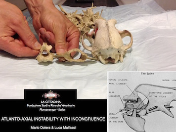
Atlantoaxial instability is a surgical condition frequently described in toy breed’s dogs. The various correction techniques have been described in veterinary literature. The aim of this study was to describe MRI-CT imaging findings and a novel surgical technique for stabilisation of atlantoaxial instability associated with incongruence of atlantoaxial joint in dogs.
Methods
Five dogs with a body weight of 2 kg or less with atlantoaxial instability associated with articular incongruence were studied. During clinical examination all dogs showed tetraparesis and ataxia. At CT and MRI, the articular surface of the atlas was larger than the articular surface of the axis, a laxity of all atlantoaxial ligaments was evident, so during the head and neck flexion the axis was dislocated dorsally and cranially into the spinal canal. Spinal cord showed a chronic compression with focal hyperintensity in seq TSE T2W and various degrees of syringomyelia. For surgery, the anaesthetised dogs were positioned in ventral recumbency with the head extended on the neck with slight linear traction applied at maxillary canine teeth. The reduction of atlantoaxial subluxation was verified through fluoroscopy. A standard dorsal approach to the atlas and axis was applied. Two 2.7 mm self tapping cortical screws were inserted on atlas winds on each side in a dorsal-ventral direction. Three 2.0 mm self tapping cortical screws were inserted transversally in the dorsal process of the axis. All screws were fused with polymethyl methacrylate. Serial clinical and imaging controls were provided.
Results
No intra-operative complications were observed. Functional improvement occurred in all dogs. Serial CT examinations showed a stable reduction of dorsal and cranial axis dislocation with persistence of a residual mild spinal compression due to the different size of the articular surfaces of atlas and axis.
Conclusion
Conclusion
Several means for stabilisation of atlantoaxial dislocation are described in veterinary literature. Standard treatment consists in ventral arthrodesis with cross pinning, transarticular lag screw fixation or vertebral plating. This technique requires atlantoaxial joint congruence. Otherwise the means of synthesis can injure the spinal cord. Dorsal techniques utilise various non-absorbable suture materials or devices such as Kishigami Tension Band. However, cranial dislocation or impingement of the axis on spinal cord are not adequately counteracted. The technique that we describe is simple and safe and can be useful in surgical treatment in dogs with atlantoaxial dislocation and articular incongruence, a condition resembling atlantoaxial invagination described in human medicine.