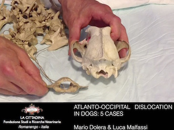
ATLANTO-OCCIPITAL DISLOCATION IN 4 DOGS: MRI FINDINGS AND SURGICAL TREATMENT
Atlanto-occipital dislocation can occur in dogs following vehicle trauma. Various surgical techniques have been proposed, sometimes with disappointing results. Aim of this work is to propose a new surgical technique for atlanto-occipital dislocation in dogs.
Four dogs suffering from atlanto-occipital dislocation were considered.
All dogs exhibit varying degrees of quadriplegia and dyspnea. Head and neck 1.5T MRI showed atlanto-occipital dislocation, with disruption of atlanto-occipital ligaments and spinal cord compression. Surgery was performed immediately. Each patient, anesthetized and mechanically ventilated, was placed in sternal recumbency. Four surgical accesses were carried out at the zygomatic processes and at the atlas wings on each side. Once these structures had been exposed, a 2-mm diameter hole was drilled in each atlas wing 5 mm caudal to the cranial margin and 10 mm medially to the margin. A nylon monofilament was inserted in the hole and an O-shaped ligature was carried out externally to the skin through the ipsilateral zygomatic arch. Once the anatomical reduction of the dislocation was controlled by fluoroscopy imaging, the surgical openings were sutured. Two weeks after surgery patients regained ambulatory status. Ligatures were removed after 2 months. Gentle manipulations aimed to verify the atlanto-occipital stability; a slight reduction in the physiological excursion in flexion and extension was observed. On follow up examinations (up to 24 months) all dogs were found normal.
Surgical technique that we developed is safe and simple to perform if compared to other described in veterinary literature. However, the limited number of cases requires further studies.