ACVIM Convention – Indianapolis, USA
4-6 june 2015


CANINE PERIPHERAL NERVE SHEATH TUMORS: CLINICAL ASPECTS, MAGNETIC RESONANCE IMAGING FINDINGS AND COMPARISON OF PALLIATION, SURGERY AND STEREOTACTIC RADIOTHERAPY
No updates for canine peripheral nerve sheaths tumor (PNST) appeared in recent literature. The aim of this study was to evaluate the correlation between clinical aspects and MRI findings of tumors involving a major peripheral nerve, plexus or root and to determine the survival time in dogs treated with palliation, surgery or stereotactic radiotherapy (SRT). Records of dogs with PNST evaluated from 2000 to 2014 were reviewed to determine signalment, duration of clinical signs, neurological examination, MRI features, treatment option (palliation, surgery, stereotactic hypo-fractionated radiotherapy). Time to first event, survival times and statistical differences across categories were calculated by the Kaplan-Meier product limit method and log-rank test. Forty-seven dogs (median age 9 years, male:female ratio 1.76) were included, with Labrador retriever over represented (17%). Roots lesions were the most frequent (46.8%), with C5-T1, V nerve and left side more involved (25.5%, 19.1% and 61.7%). Presenting signs were lameness, paresis and pain. Mean duration of clinical signs was 90 days. MRI findings comprises increased diameter, hyperintense and contrast enhancing nerve roots (57.1%), plexus or peripheral nerve (42.9%), focal hypomiotropy and muscle hyperintensity (73%). The time to first event was 30 days after surgery and 240 days after SRT. Overall mean survival was 97, 144 and 371 days with palliation, surgery and SRT. A predilection for Labrador retriever is observed. Comparing our results with published data, SRT seem to promise better results than palliation or surgery and warrant further evaluation.
DEFINITIVE HIGH-DOSE HYPO-FRACTIONATED STEREOTACTIC BRAIN-SPARING IRRADIATION OF STAGE IV CANINE NASAL TUMORS: A FEASIBILITY STUDY AND FIRST CLINICAL EXPERIENCES
The prognosis for canine nasal tumors with intracranial extension is poor with an expected survival of 1 month with palliation and 6.7 months with irradiation. However, studies regarding stage IV nasal tumors treated with brain-sparing irradiation techniques are lacking. The aim of this prospective study was to evaluate feasibility and efficacy of definitive intent stereotactic radiotherapy in dogs with nasal tumors with massive intracranial extension. Seven dogs with stage IV nasal tumors were treated with high-dose hypo-fractionated stereotactic radiotherapy with VMAT technique. Dose prescriptions were 32-36 Gy in four consecutive-day fractions to the gross tumor and 30 Gy to lymphatics. Adjuvant treatment included carboplatin. Serial clinical and CT/MRI examination were performed. Disease control and toxicity effects were evaluated according to RECIST and VRTOG criteria. Median survival time (MST) was evaluated using Kaplan-Meier curves. Six carcinoma and 1 sarcoma were treated. Prescription goals were obtained in four cases with V95%>95% and V107%>2% whereas in 3 dogs V95%=86-90% was accepted to limit maximum brain punctual dose <27 Gy. Two partial response and 5 complete responses were obtained. MST was 9 months. One grade II late brain radiotoxicity and two brain ascending infections were observed. Relapse pathways involves diffuse meningeal and sphenoid invasion. The initial experiences with the RT regimen adopted indicate a feasibility and effectiveness in modified stage IV nasal tumors. The relapse pathways observed suggest to evaluate alternative adjuvant treatment in dogs treated with stereotactic radiotherapy.
DEFINITIVE HIGH-DOSE HYPO-FRACTIONATED TOTAL PELVIC IRRADIATION WITH SIMULTANEOUS BOOST IN CANINE URINARY TRANSITIONAL CELL CARCINOMA: A FEASIBILITY STUDY AND FIRST CLINICAL EXPERIENCES
The lower urinary tract transitional cell carcinoma (CCT) poses challenge in order to the appropriate radiotherapy (RT) regimen. Organs at risk (OARs) within the irradiation field (ureters, rectum) are the limiting factors in dose escalation. The primary aim of this study was to evaluate the technical feasibility of high-dose hypo-fractionated dynamic IMRT in non-resectable lower urinary CCT affected dogs. The secondary goal was to evaluate the toxicity and the efficacy of the RT regimens. Three dogs with lower urinary tract CCT were treated with definitive high-dose hypo-fractionated RT with volumetric modulates arc therapy (VMAT) technique. The volume treatment definition include the gross tumor (GTV), the PTV1 (GTV+3mm), lymphatics (PTV2), the entire bladder, prostate in males and uretra (PTV3), the entire pelvis except the rectal volume (PTV4). Dose prescriptions were 40 Gy to PTV1, 38 Gy to PTV2, 34 Gy to the PTV3, 30 Gy to the PTV4, in 6 fractions on alternate days. A piroxicam was subministered to all dogs. Serial clinical and CT/MRI examination were performed. Disease control and toxicity effects were evaluated according to RECIST and VRTOG criteria. Three T2N0M0 urinary tract CCT were treated. Prescription goals were obtained in all three cases with V95%>95% and V107%>2%. During follow-up (mean 6 months) one partial response and two complete responses were obtained. Two grade I cystitis were developed. Non rectal toxicity was recognized. The initial experiences with the RT regimen adopted indicate a feasibility and effectiveness in lower urinary CCT.
PERIPHERAL GLYCAEMIA IN DOGS WITH LIMB THROMBOSIS: A PROSPECTIVE STUDY
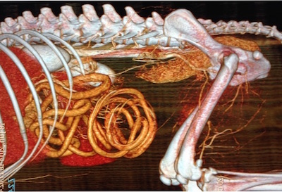
The aim of this study was to document the peripheral glycaemia variations in hypoperfused limbs of patients affected by Magnetic Resonance Imaging (MRI)-confirmed arterial thrombosis. Eleven dogs were recruited. Inclusion criteria were a clinical examination supportive of limb hypoperfusion and availability of blood cell count, biochemical profile and urine analyses. Two blood samples were sampled, one from the affected limb and one from a healty limb. Plasmatic glycaemia was measured using an automated glucose analyser. All the patients underwent a total body MRI that provided the final diagnosis. The thrombus was located: in the abdominal aorta (7/11), in the subclavian artery (1/11), in the axillary artery (1/11), in the iliac arteries (2/11). Of the total abdominal aortic thrombosis, 3/7 involved also the internal iliac arteries, 2/7 the external ones and 2/7 both. The extent of the thrombosis was classified as grade 1 when the greatest portion of the thrombus did not reach half of the vessel lumen (1/11); grade 2 when the greatest portion of the thrombus was between 1/2 and 2/3 of the vessel lumen (7/11); grade 3 when the thrombus exceded 2/3 of the lumen (3/11). A substantial decrease in peripheral glycaemia values was found in sampling arising from the affected limbs. Comparing affected limbs values with healthy limbs measurements from the same patient, the reduction was found from 17.65% to 34.41%. Accounting only the grade 3 scored patients, the percentage of reduction was found up to the 28.34%.
SURGICAL STABILIZATION OF CANINE LUMBOSACRAL SPINE WITH STOP-SCREWS AND ILIAC WINGS SCREWS
Surgical stabilization of canine lumbosacral spine can be challenging. The aim of this research was to evaluate two surgical techniques to achieve lumbosacral stabilization in dogs either with normal or transitional vertebrae. Lumbosacral instability and degenerative stenosis were evaluated by dynamic Computed Tomography (CT) and Magnetic Resonance Imaging (MRI). In dogs with normal vertebrae two 4.5 mm screws were bicortically inserted in S1 with the heads behind the caudal articular process of L7 to prevent the extension of the lumbosacral joint; if ventral listhesis of S1 was evident, the head of the screws were augmented by methyl methacrylate. In dogs with transitional vertebrae, two 4.5 mm screws were inserted in the iliac wings, two 3.5 mm screws were inserted in the spinous process of L6 and L7; the emerging screws were embedded in methyl methacrylate after flexion of the lumbosacral spine. In cases of residual radicular compression, dorsal laminectomy and partial discectomy were accomplished. Serial clinical and imaging follow-up examinations were performed. Twenty-two large breed dogs were enrolled. In 14 dogs stop-screws (in 4 augmented) and in 8 dogs iliac wings screws were inserted. 2 dogs required additional decompression. During a mean follow-up of 36 months, clinical examination and imaging reveals amelioration of presenting complaints and reduction of radicular compression, with no surgical complications. Stop-screws and iliac wings technique are effective methods to obtain stabilization and indirect decompression of the lumbosacral joint. Comparing with other described surgical procedures, our obtained results are better but with lesser complications.


